


Case Presentation:
Degenerative Disk Disease - Case 1
History & Physical
- 71-year-old man with a long history of low back pain, however, with increasing vehemence over the past year with 10/10 intensity on Visual Analogue Scale and the development of pain radiating in the posterior aspect of both thighs.
- His neurological examination was normal with no weakness, or numbness of the lower extremities, however, significant pain with range of motion of the lumbar spine.
Imaging
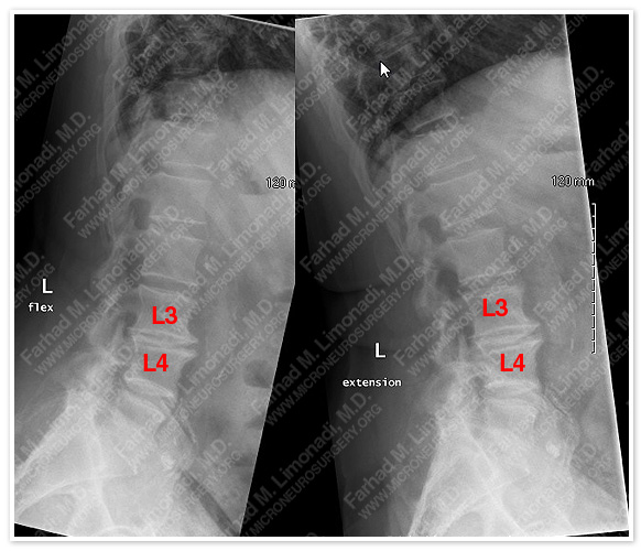
Plain flexion-extension x-rays of the lumbar spine show no instability, but instead significant degenerative disk disease at L3-L4 level.
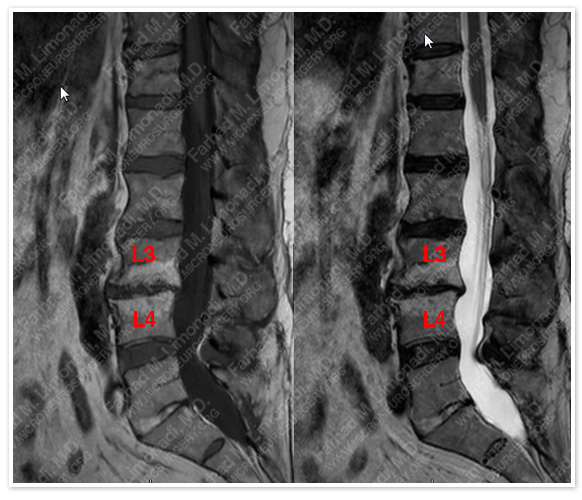
MRI scan of the lumbar spine confirms significant degenerative disk disease at L3-L4 level (Modic type II changes at L3-L4 level).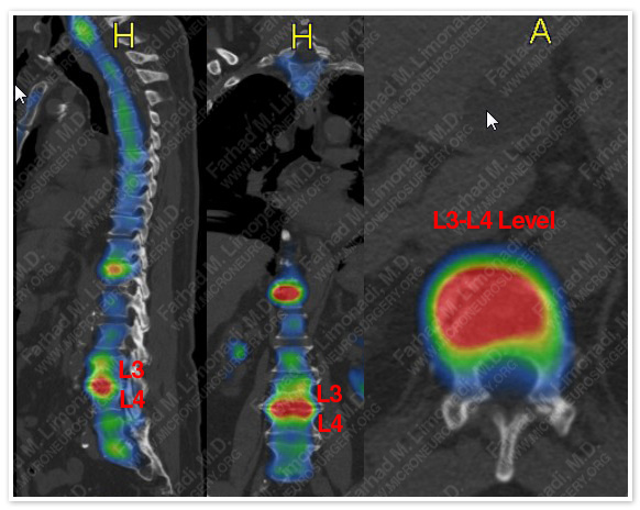
CT SPECT of the lumbar spine confirms significant degenerative disk disease at L3-L4 level (MDP uptake at L3-L4 level).
Surgical Procedure
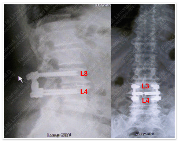
- After a comprehensive battery of conservative measures, he underwent PLIF (Posterior Lumbar Inter-body Fusion) at L3-L4 level.
Post-op Course
- He did well postoperatively with immediate improvement of his function and was discharged home. Patient’s low back pain and thigh pain completely resolved postoperatively.