


History and Physical
- 78-year-old right-handed lady, with new onset of facial pain 6 month before presentation. Her pain was described as an electric shock and jolt in the right V2 and V3 distribution (cheek and chin area) with intensity of 10/10 on Visual Analogue scale, initially lasting a few seconds, but by presentation lasting 5-10 minutes each time. Her pain was triggered by application of water or cream to face, brushing her teeth or chewing. She was appropriately initially treated with Tegretol and Trileptal, and Neurontin. Despite initial benefit, she became refractory to these medications. She was seen in the emergency room multiple times due to pain as the result.
- On examination she had persistent numbness in the right facial pain in the distribution of pain as well as mild facial droop.
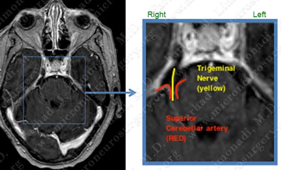
MRI of her brain shows branches of superior cerebellar artery pressing on Trigeminal Nerve.
- Having been correctly diagnosed with trigeminal neuralgia, and also refractory to proper medical management , she underwent microvascular decompression of trigeminal nerve by a right retrosigmoid craniectomy with intra-operative neurophysiological monitoring and stereotactic computer aided navigation.
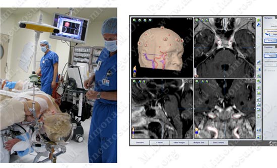
Patient is properly positioned on the operating table. Computer navigation and stereotaxy is utilized to co-register her brain coordinates with the computer system.
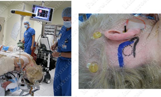
Critical structures such as transverse and sigmoid sinuses are mapped and marked (blue on right picture). A key hole approach is planned (dotted circle). Brain auditory evoked response microphone is inserted in her ear canal.
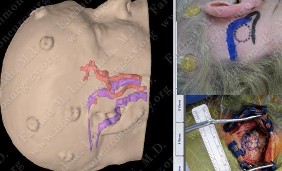
A small incision behind the ear exposes the bony anatomy for a key hole approach.
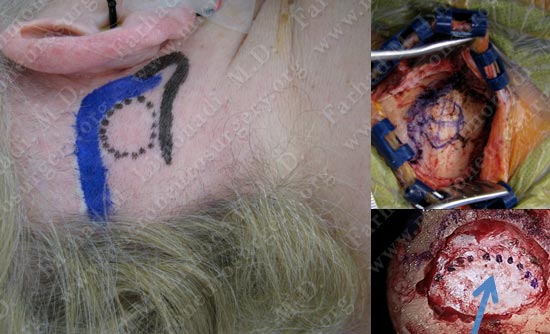
After bony exposure the dura is exposed and opened carefully.
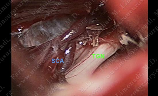
Trigeminal nerve (TGN) is distorted and compressed by a pulsating superior cerebellar artery (SCA) and its branches.
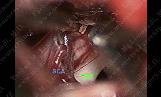
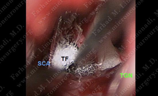
Pulsating superior cerebellar artery (SCA) is being dissected off Trigeminal nerve (TGN) using micro-instruments under the high power magnification of microscope.
- Patient did well post-operatively with complete resolution of his facial pain. She was discharged home and returned to full function.
- She remained pain free and was successfully weaned off her pain medication.