


Case Presentation:
Meningioma - Case 14
History & Physical
- 60-year-old lady with new onset of seizure while jogging and significant facial injuries as the result.
- On examination, she had no focal neurological deficits.
Imaging
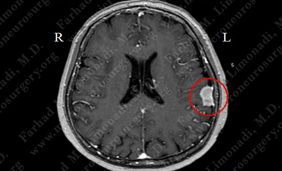
MRI scan of the patient's brain showed a left parietal tumor arising from the dura (covering of brain).
Computer Navigation
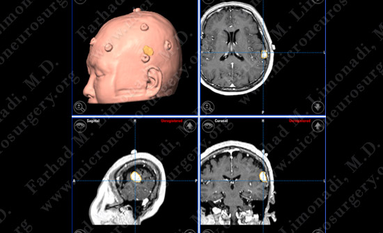
Stereotaxy and computer navigation was utilized to localize the tumor precisely. Tumor is outlined in yellow.
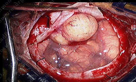
Surgical Protection
- Small circular bone flap is elevated using computer localization. The dura overlying the tumor excised and the tumor revealed.
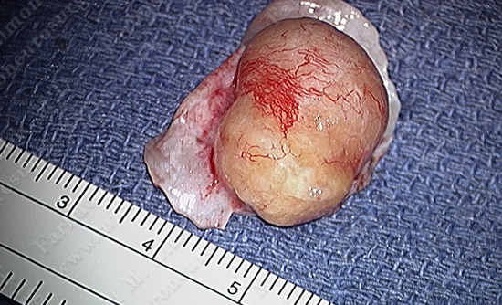
Pathology
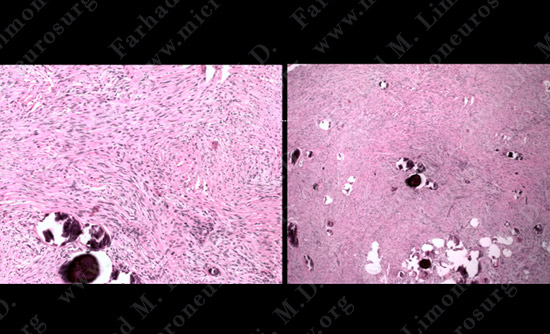
H&E slides of tumor
The pathology of the tumor confirmed diagnosis of meningioma.
Post-op Imaging
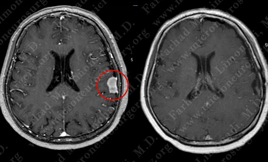
Before Operation After Operation
Post-op MRI shows complete resection of the tumor with no injury to surrounding neurovascular structures.